Online High School Biology: Draw and Revise a Cell Model
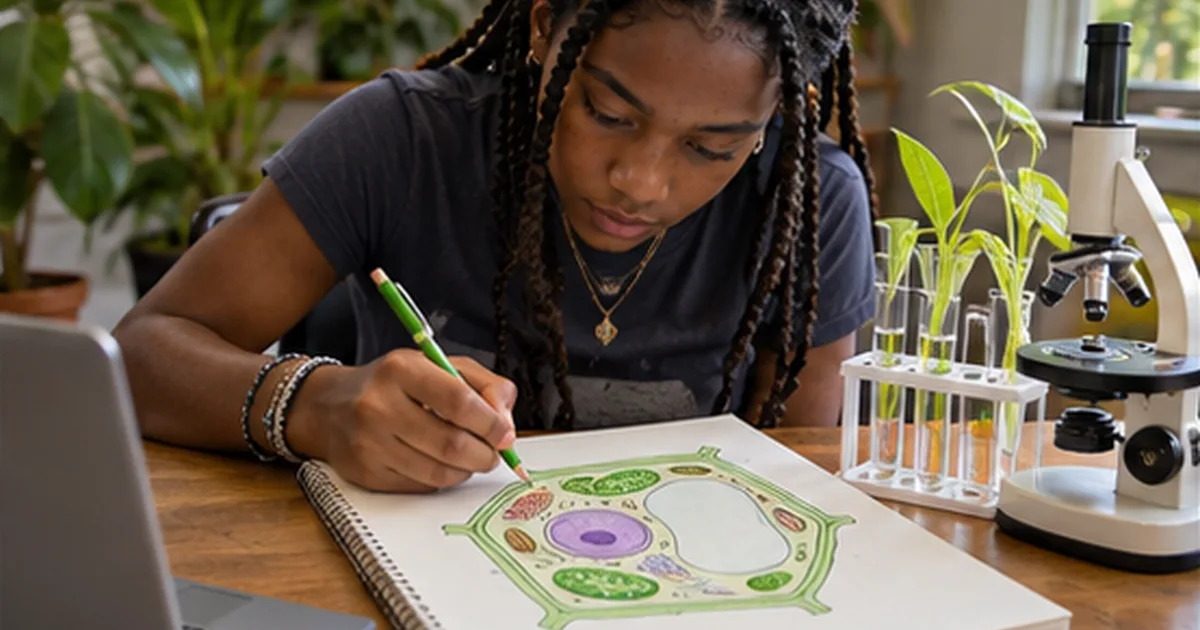
Draw the boundary of a plant cell twice: once for the cell wall and once for the membrane. Then explain what each boundary does. If the explanation is harder than the drawing, the model has already identified a useful question. Revisit the drawing after feedback and make each new label explain a function or relationship.
Roovet Academy provides homeschool information and offers a course catalog that includes science subjects. Explore available biology courses at online high school classes to see how cell modeling might fit into a self-directed learning plan.
Why Model a Plant Cell?
Modeling is more than drawing—it’s a form of scientific thinking. When students create a visual representation of a plant cell, they must make decisions about scale, component placement, and functional relationships. This forces them to engage with the material actively rather than passively.
A plant cell differs from an animal cell in several key ways: it has a rigid cell wall, large central vacuole, and chloroplasts. Accurately depicting these features requires understanding their roles. For example, the cell wall isn’t just a boundary—it provides structural support and protection. The membrane inside regulates what enters and exits. Students who merely label parts on a worksheet may miss these distinctions. But when they draw and revise, they confront gaps in understanding.
Step-by-Step: Drawing the Initial Model
Start with a clear objective: draw a plant cell, labeling at least eight organelles and explaining two functions each for the cell membrane and cell wall. While magnification levels like 400x may vary by microscope, the focus should be on accurate representation of structures rather than a specific technical setting.
Take the case of Maria, a 10th-grade student in an online biology course. Her first attempt shows a large rectangular cell—correctly shaped due to the cell wall. She includes the nucleus, mitochondria, and chloroplasts. Her labels are neat, but her explanation of the cell membrane says only “keeps things in.”
This is a common starting point. Students often conflate the membrane and wall or oversimplify their roles. Maria’s next step isn’t just redrawing—it’s revising based on feedback.
Providing Effective Feedback
Constructive critique is essential. Instructors or learning coaches should focus on clarity, accuracy, and reasoning. For Maria, feedback might highlight:
- The cell membrane is selectively permeable—explain how it controls movement of water and nutrients.
- The cell wall is made of cellulose and prevents bursting in hypotonic environments—connect structure to function.
- Chloroplasts should show internal thylakoids if possible, indicating photosynthetic capacity.
Feedback should also guide presentation. For example, suggest using color coding: green for chloroplasts, blue for vacuoles, and red outlines for the membrane. This visual strategy reinforces learning.
Revising After Critique: Maria’s Second Draft
Maria revises her model with clearer detail. She adds:
- A detailed cell membrane with embedded proteins to illustrate selective transport.
- A thick, layered cell wall labeled as cellulose, with a note that it maintains turgor pressure.
- An enlarged central vacuole pushing the cytoplasm toward the edges, showing its role in storage and structure.
Her written explanation now reads: “The cell membrane uses protein channels to allow water and ions in while blocking harmful substances. The cell wall, outside the membrane, gives rigidity so the plant doesn’t wilt.”
This revision shows conceptual growth. She’s no longer listing parts—she’s explaining systems.
Common Misconceptions to Address
Even with guidance, students often misrepresent key features. Watch for these errors:
- Confusing membrane and wall: Some draw the membrane outside the wall. Emphasize sequence: wall (outermost), then membrane just beneath.
- Incorrect vacuole size: In mature plant cells, the central vacuole occupies up to 90% of volume. Small vacuoles in drawings suggest misunderstanding.
- Misplaced chloroplasts: They should be scattered in the cytoplasm, not clustered. Their distribution maximizes light absorption.
- Oversimplified nucleus: Include nucleolus and nuclear pores to show RNA export and regulation.
Correcting these in revision strengthens accuracy. For example, when James, another student, drew tiny vacuoles scattered throughout his cell, feedback prompted him to research turgor pressure. His second version shows a single large vacuole pressing against the membrane, with a caption: “This pressure keeps the plant upright.”
From Drawing to Deeper Questions
A strong model should prompt inquiry. After completing her revised drawing, Maria is asked: “What would happen if the cell wall were removed?”
She reasons: “Without the wall, the cell would rely only on the membrane. In a hypotonic solution, it might swell and burst—like an animal cell.” This shows she understands osmosis and structural differences.
This kind of application is critical. It moves learning from “what” to “why” and “what if.” Online courses that incorporate open-ended questions after modeling tasks deepen comprehension.
Using Digital Tools Effectively
Many students use digital platforms to create and revise models. A simple drawing app allows layering—students can place the wall, then membrane, then organelles. Some use slide-based tools to show progression: first draft, feedback, second draft.
However, analog methods work too. Pencil and paper allow easy erasing and redrawing. The medium matters less than the process of iteration.
What’s essential is that revisions are visible and justified. Students should annotate changes: “Added protein channels in membrane to show active transport.” This builds scientific communication skills.
Assessing the Final Model
Evaluation should focus on three areas:
- Accuracy: Are organelles correctly placed and labeled?
- Explanation: Does the student describe how membrane and wall function?
- Revision: Can they articulate what changed and why?
For instance, Chloe submits a final model with a detailed legend. She writes: “I originally drew the cell wall as thin. After feedback, I thickened it and added cellulose fibers. I also learned that plasmodesmata connect plant cells through the wall—so I included those.”
This reflection demonstrates metacognition—thinking about thinking. It’s a hallmark of deep learning.
Connecting to Broader Concepts
Cell modeling isn’t isolated. It’s a gateway to topics like osmosis, photosynthesis, and plant anatomy.
After modeling, students can explore:
- How root hair cells differ in structure to increase surface area.
- Why guard cells have uneven walls to enable stomatal opening.
- How turgor pressure affects seedling growth.
These extensions help students see cells not as static diagrams but as dynamic systems.
Supporting Independent Learners
In online settings, students often work without immediate peer interaction. Parents or learning coaches can support by asking targeted questions:
- “Can you point to where the cell membrane is?”
- “How does the vacuole affect the position of other organelles?”
- “What would happen if chloroplasts were missing?”
These prompts encourage self-assessment. They also prepare students for written or oral defense of their models.
One challenge in homeschool settings is ensuring scientific accuracy without formal lab access. But modeling compensates—students learn through deliberate representation and correction. A well-revised cell diagram can demonstrate mastery equivalent to traditional lab reports.
Preparing for Advanced Biology
Skills developed in cell modeling—observation, critique, revision—are foundational for AP Biology or college-level courses. For example, understanding membrane structure is essential when studying signal transduction or neuron function.
Students who practice explaining the lipid bilayer and transport proteins early are better prepared for complex topics like facilitated diffusion or the sodium-potassium pump.
Consider Ethan, who struggled initially with organelle functions. After revising his plant cell model three times, he began to see patterns: membranes regulate movement, organelles compartmentalize tasks. This systems-thinking approach helped him later understand cellular respiration in mitochondria.
Conclusion
Drawing and revising a plant cell model is more than an art project—it’s a scientific practice. It teaches students to observe, represent, critique, and improve. When integrated into an online biology curriculum, this process fosters deep understanding of cellular structure and function.
Through concrete examples like Maria’s and James’s revisions, we see how feedback transforms superficial drawings into meaningful learning tools. By focusing on the cell membrane and wall—not just as parts, but as functional systems—students build a foundation for future study.
Families interested in structured, project-based science learning can find resources at homeschool curriculum support to guide these experiences effectively.
Explore Roovet Academy’s biology course.